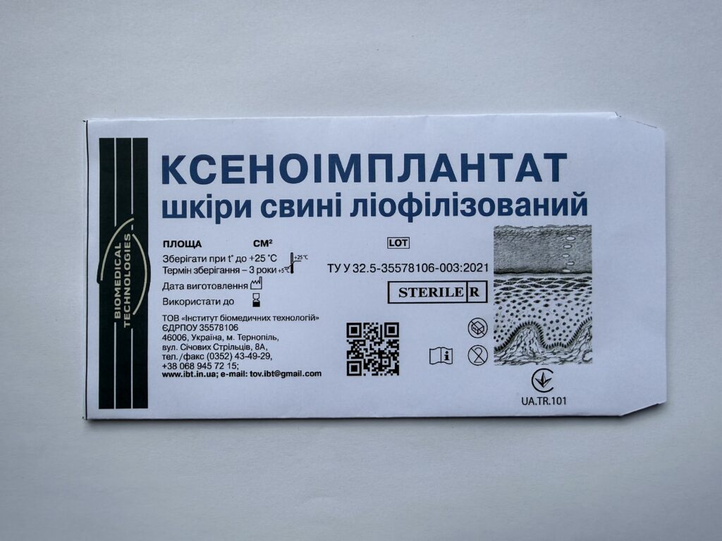
Based on the patented invention of Professor V.V. Bihuniak, a methodology for obtaining, cryopreservation, and lyophilization of porcine skin as an ideal skin substitute has been developed. Under his supervision, a bank of lyophilized xenoimplants was established, which annually, since 1996, produces and delivers up to 1 million cm² of lyophilized xenoimplants to medical institutions across Ukraine, reliably supplying all burn centers and healthcare facilities throughout the country in both scheduled and emergency cases.
Combustiologists of Ukraine have demonstrated in practice that superficial burns covered with xenoimplants heal significantly faster, while for patients with deep burns and extensive skin damage, the use of xenoskin represents their only chance of survival. Xenoimplants are also effectively used in the treatment of donor site wounds, degloving wounds, trophic ulcers, and wounds following cryodestruction of scars.
Characteristic
Lyophilized xenoimplants with an area of 100–150–200–250–300 cm², a thickness of 0.3–0.4 mm, sterile, packaged in pouches, require no special transportation conditions.
Indications
Lyophilized porcine skin xenoimplants are used as temporary skin substitutes in the treatment of burn wounds (grade I–IIAB–III), donor site wounds, degloving wounds, trophic ulcers, and wounds following cryodestruction of scars.
When xenoimplants are used in the complex treatment of burn patients, the general condition of the patients improves, as does their sleep and appetite; body temperature normalizes, the deficit in homeostasis parameters decreases, blood serum toxicity levels drop, epithelization of superficial burns accelerates, along with marginal and island epithelization of deep burns and donor site wounds, resulting in a 23% reduction in the area of granulating wounds. The duration of inpatient treatment is shortened by 16–18 days, and mortality among severely burned patients is reduced by 30%.
The number of lyophilized xenoimplants required to close wounds depends on the area and depth of the burn wounds and the age of the patient.
Application Guidelines
For superficial burns (grade I–IIA)
After the patient is admitted to hospital and hemodynamics are stabilized, wound toilet is performed under anesthesia in a clean dressing room or operating theater, and the wounds are covered with lyophilized xenoimplants. Epithelization of wounds under lyophilized xenoimplants occurs within 10–12 days.
In grade IIA burns, the dermal layer partially dies (sebaceous and sweat glands and their ducts remain viable), which creates conditions for wound suppuration caused by the development of opportunistic microflora in the necrotically altered superficial skin tissues.
To achieve conditions for active island and marginal epithelization through preserved skin derivatives, it is necessary to timely remove necrotic tissue and combat wound infection. Therefore, patients with grade IIA burns should undergo sequential (superficial) necrectomy in the early period after injury (within 2–3 days), and wounds should be covered with lyophilized xenoimplants. This prevents the development of burn disease and associated complications, helps avoid frequent painful dressing changes, accelerates recovery, and prevents the formation of pathological scars.
The applied xenoimplants adhere closely to the wound, leading to an improvement in the general condition of patients, a significant reduction or elimination of pain syndrome, and normalization of body temperature.
The first dressing change after tangential and sequential necrectomy with xenoplasty is performed the following day. Subsequent dressing changes are carried out depending on the nature of implant engraftment — daily or every other day.
If hematomas are detected under the xenoimplants or if seropurulent discharge accumulates, they are removed, a repeat wound toilet is performed, and new xenoimplants or wet-to-dry dressings are applied. On days 8–9 after injury, drying of the xenoimplants at the wound edges, their rejection, and epithelization of the wound surface beneath them are observed. In other areas of the wound, xenoimplants remain firmly fixed to the underlying tissues.
On days 11–12, the transplants consolidate, dry out, and fall off. The wound surface becomes covered with a well-developed epithelial regenerate.
Thus, the use of lyophilized xenoimplants in the treatment of superficial burns allows the duration of inpatient treatment to be shortened by 6–8 days and reduces the risk of hypertrophic and keloid scar formation by 38%.
Given that the proportion of patients with grade I–IIA burns reaches 70% of all burn patients, the use of lyophilized xenoimplants makes it possible to avoid daily painful dressing changes, promotes faster wound healing, prevents suppuration, and allows the burn disease to progress without loss of proteins, water, and electrolytes. Since the cost of purchasing lyophilized xenoimplants is lower than that of ointments, bandages, solutions, anesthesia medications, etc., it can be stated that this treatment method has not only a pronounced clinical but also an economic effect.
For deep burns (grade IIB–III)
Treatment of deep burns can be carried out both with and without early necrectomy.
With the use of early necrectomy
The ultimate goal of local treatment of deep burns is the surgical restoration of lost skin cover. Early surgical interventions best correspond to the principles of preventive surgery. In this case, necrotic tissues are removed in a single stage, either tangentially or suprafascially, over an area of up to 10–15% of the body surface. The resulting wounds are temporarily covered with lyophilized xenoimplants, which are removed after 2–3 days, followed by additional necrectomy and closure of the wounds with autodermotransplants.
In the presence of large-area wounds, repeat operations are performed 2–3 days after the first autoplasty. The use of lyophilized xenoimplants makes it possible to increase the area of single-stage excision of necrotic tissues, reduce the traumatic nature of interventions, identify areas of incomplete removal of necrotic tissues, and creates conditions for faster compensation of postoperative homeostasis disturbances.
Additional removal of non-viable tissues promotes better engraftment of autodermotransplants. The use of early necrectomy with xenodermoplasty prevents progressive intoxication from the injury site and the development of wound infection, reduces the possibility of further development of burn disease, and leads to restoration of skin cover in the shortest possible time.
Treatment of deep burns without early necrectomy
Treatment of deep burns without early necrectomy is carried out in patients with purulent wounds who have been delivered from evacuation stages with delay, as well as in patients with a severe course of burn disease and concomitant conditions that limit the possibility of early necrectomy.
The main goal of treatment for burn patients is the fastest possible preparation of burn wounds for autodermoplasty. Spontaneous rejection of necrotic tissues lasts 4–5 weeks. The presence of burn eschar on the burn wound, especially moist eschar, and the vegetation of microflora in the wound (most often in the form of associations of gram-positive and gram-negative flora) frequently leads to complications of burn disease.
After chemical or staged necrectomy and cleansing of deep burn wounds, autodermoplasty is performed. During autodermoplasty, wounds not yet covered with autodermotransplants, donor site wounds, and perforated autoskin flaps are covered with lyophilized xenoimplants.
Xenoimplants can remain fixed on wounds for up to 2.5–3.5 weeks. The use of lyophilized xenoimplants during autodermoplasties leads to a reduction in pain intensity, a decrease in plasma loss, and a lower frequency of wound suppuration.
During this time, granulation tissue matures fully under the xenoimplants, in which cells of histiogenic and hematogenous origin (fibroblasts and histiocytes) are observed.
Electron microscopy of fibroblast lineage cells reveals hypertrophy of protein synthesis and energy metabolism structures.
Simultaneously with the formation of granulation tissue, epithelization of the wound surface proceeds more actively; alongside marginal epithelization, local spread of epithelium is observed in the form of broad cellular outgrowths from preserved skin derivatives.
This leads to a reduction in wound area due to the absence of secondary wound deepening and necroses, as well as enhanced marginal and island epithelization of deep burn wounds under xenoimplants.
After removal of the xenoimplants, the wounds are ready for autodermoplasty.
Closure of Donor Site Wounds
Xenoimplants are effectively used for the treatment of donor site wounds. This eliminates the need for dressing changes. Epithelization of donor site wounds under xenoimplants occurs on days 6–8.
Thus, the use of xenoimplants for closure of donor site wounds leads to faster epithelization (by (4±1) days), which makes it possible, when necessary, to harvest autotransplants more quickly for repeat grafting.
Closure of Perforated Autodermotransplants
Perforated autodermotransplants on wounds can be covered with xenoimplants. Xenoimplants should not be removed during dressing changes; epithelization of wounds in the cells of the autograft proceeds under the xenoimplants.
After complete epithelization of wounds between the strands of the perforated autotransplant, the xenoimplants become dry and fall off.
Treatment of Degloving Wounds and Trophic Ulcers
Clean degloving wounds and trophic ulcers are covered with lyophilized xenoimplants. Under engrafted xenoimplants, a reduction in the inflammatory process and activation of marginal and island epithelization are observed, leading to spontaneous wound healing.
For large wounds, the xenoflaps should be replaced with autodermotransplants on days 4–6 (during this period, local blood circulation in the wound is at its best).
Treatment of Wounds Following Cryodestruction of Scars
On the second day after cryodestruction, the affected area (blister with serous content and surrounding skin) is irrigated with antiseptic solutions, the blister with serous content is removed, the wound is dried with sterile gauze, and a lyophilized xenoimplant is applied.
On days 6–7–8 after the procedure, drying of the xenoimplant at the wound edges, its rejection, and complete epithelization of the wound surface beneath it are observed. The duration of epithelization depends on the area of the wound formed after cryodestruction.
Storage conditions
Xenoimplants should be stored at a temperature of +5 °C to +25 °C for up to 3 years. They are lightweight, with one package weighing 70–120 g.
Scientific Evidence of Cell Viability Preservation in the Production of Xenoimplants
Microscopic examination of lyophilized skin revealed no signs of autolytic or necrobiotic changes or profound structural alterations in the epidermis or papillary layer of the dermis. The nuclei and cytoplasm of epidermiocytes and fibroblasts are well preserved in the vast majority of cells, with pyknosis and vacuolization present only in a small proportion. Cell membrane definition is maintained, and no detachment of the epidermis from the dermis is observed. Collagen fibers of the dermis are contoured in the overwhelming majority of specimens, form a network, and are loosely arranged in the papillary layer. In the deeper portions of the dermis, edema and homogenization of individual areas of collagen fibers are noted, and in some cases their fragmentation.
Van Gieson picrofuchsin staining reveals that the collagen fibers are predominantly stained fuchsinophilically in an intense red color. The portion of the elastic network that forms part of the connective tissue framework is represented by clearly contoured thin elastic fibers.
Electron microscopic examination at all observation time points clearly reveals plasma membranes and intercellular contacts of the germinative layer of the epidermis. Euchromatin predominates in the nuclei, indicating preserved functional activity of the nuclear apparatus of epitheliocytes. Among the cells of the papillary layer of the dermis, fibroblasts predominate, characterized by a well-developed protein synthesis apparatus and minor mitochondrial damage. The contours of the nuclei, in which numerous granules of the ribosomal type are observed, are clearly defined.
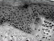
figure 1
Preserved layered arrangement of epidermoсytes relative to a distinct basal membrane. Basophilic nuclei, vacuolization of the cytoplasm of individual cells.
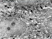
figure 1а
Electron microscopic organization of epidermoсytes of the spinous layer. Preserved desmosomal contacts, moderately widened intercellular spaces. Minor invaginations of the karyolemma and homogeneous karyoplasm of the nuclei, destruction of individual organelles.