Dental membranes
Lyophilized porcine tissue xenoimplants (dermis, peritoneum, pericardium)
Sizes:
- S: 2×3 cm
- L: 3×4 cm
Thickness:
- Dermis — 0.4 mm; 0.7 mm; 1.0 mm; 1.5 mm.
- Peritoneum — 0.3 mm; 0.6 mm.
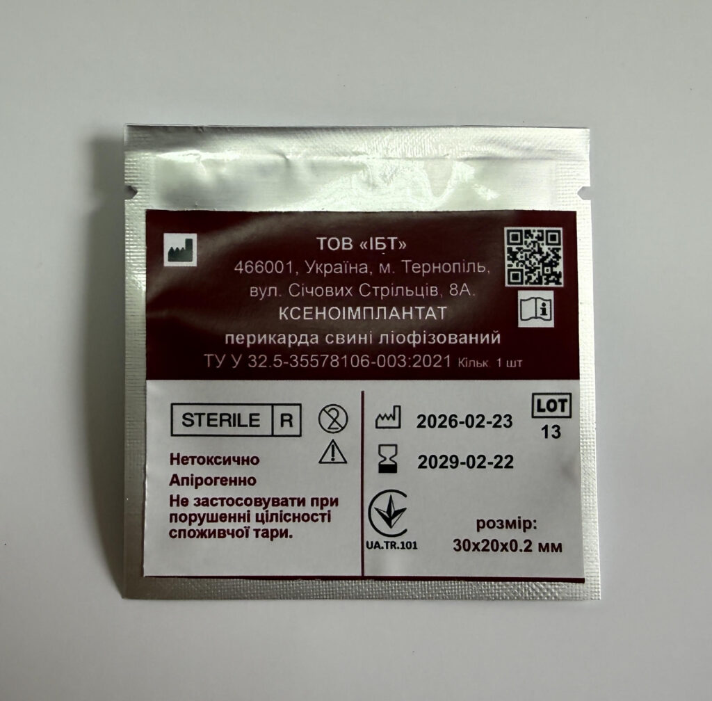
- Pericardium — 0.2 mm.
Most modern protocols for guided bone or guided tissue regeneration involve the use of membranes during surgery. They serve a barrier function, preventing the proliferation of soft tissues into the bone augmentation zone, as well as retaining the grafting material. We have conducted studies on the potential use of xenogenic tissues in dental practice. It has been demonstrated that xenoimplants of dermis, pericardium, and peritoneum, manufactured using cryopreservation and lyophilization technology, can serve as membranes for the temporary closure of mucosal wound defects in periodontology, implantology, dental and maxillofacial surgery.
Characteristic
The positive properties of membranes made from dermis, pericardium, or peritoneum include ease of use, optimal wound healing, natural biomechanical effect, and the presence of a strong multi-directional collagen network that ensures a long-lasting barrier function for 3-6 months.
Membrane properties:
- long-lasting barrier function: ~ 12-24 weeks
- natural structure and low thickness
- ease of use (can be used both in dry and moist conditions)
- high elasticity and strength
- excellent adaptation to surface contours
- no adhesion after rehydration
- rapid revascularization due to the three-dimensional structure
- good strength and tear resistance
Indications
Indications for the use of the dermis membrane:
Periodontology: Dermis can be used for guided tissue regeneration in the treatment of periodontal diseases and for covering gingival recession. The membrane can be applied in combination with the following techniques:
- coronally advanced flap
- laterally repositioned flap
- envelope technique
- tunnel technique
Implantology, dental and maxillofacial surgery:
Xenogenic tissues can also be used:
- for local augmentation of the alveolar ridge prior to implantation
- for alveolar ridge restoration before subsequent treatment
- after tooth extraction, after cystectomy and root apex resection
- in soft tissue volume augmentation procedures
- for implant coverage or in immediate or delayed implant placement into the extraction socket
Indications for the use of the peritoneum membrane:
- coverage of the implantation area
- sinus lift
- protection of the Schneiderian membrane
- closure of fenestrations
- sealing of the post-extraction socket
- prevention of alveolar ridge atrophy
- horizontal and lateral augmentation
- alveolar ridge reconstruction
- intrabony defects (1-3 walls)
- furcation defects (Class I-II)
Indications for the use of the pericardium membrane:
- coverage of the implantation area
- sinus lift
- protection of the Schneiderian membrane
- prevention of alveolar ridge atrophy
- horizontal and lateral augmentation
- alveolar ridge reconstruction
- intrabony defects (1-3 walls)
- furcation defects (Class I-II)
Contraindications:
Do not use in the presence of acute inflammatory processes in the surgical area.
Usage recommendations:
Preparation:
- the product is submerged in sterile saline solution for 5 minutes at room temperature
- the membrane is manually shaped as required
- if necessary, the membrane is trimmed instrumentally
Use of membranes without soaking in saline solution is permitted.
Basic principles of using dermis, peritoneum, and pericardium membranes:
- contact between the membrane and the periosteal bed and the mobilized mucosal flap must be ensured by placing interrupted or cross sutures
- trimming the edges of the membrane while slightly moistened prevents damage to the gingival tissue when covering the membrane with a flap
- fixation of the membrane with interrupted or cross sutures, titanium pins, or screws is required
Use
Indications for the use of cryo-lyophilized xenogenic membranes:
- for prevention of bone atrophy following tooth extraction
- for fixation of bone graft granules during jaw bone augmentation procedures
- barrier membranes serve as a reinforcing agent during dental implantation with simultaneous placement of bone granules
- for stabilization of mobile teeth in periodontal tissue diseases
Principles of working with membranes in bone augmentation:
- the membrane must completely cover the augmentation material and should therefore be slightly larger in size
- fixation of membranes is performed using titanium pins, screws, or suture material attached to fixed landmarks in the surgical wound, i.e., to the bone or periosteum
- proper technique and handling of the mucoperiosteal flap must be observed so that it can cover the membrane without tension
- the membrane must not resorb before bone formation is complete, which depends on the thickness of the xenogenic tissue used
Storage conditions
Membranes must be stored in a dry place (at a temperature of +5 °C to +25 °C).
Scientific rationale:
Preclinical and clinical studies have been conducted. Animal experiments on pigs examined the resorption times of xenogenic tissues — the dermal skin layer, peritoneum, and pericardium — following subcutaneous, subfascial, and submuscular implantation of the xenoimplant, depending on its thickness.
The amino acid and trace element composition, as well as the presence of biologically active substances (growth factors) in cryolyophilized xenogenic tissues, were investigated.
Qualitative and quantitative analysis of the trace element and amino acid composition of xenosubstrates revealed the presence of amino acids such as arginine, valine, histidine, isoleucine, leucine, lysine, threonine, phenylalanine, and others, providing insight into the biological activity of these substrates as organ preparations with pluripotent properties.
The high redox potential of the xenosubstrate, associated with the content of amino acids, oxidative enzymes (peroxidase, cytochrome oxidase), and other biologically active substances — in particular, acidic mucopolysaccharides (hyaluronic acid, chondroitin sulfuric acid, and heparin) — is further supported by a significant concentration of trace elements in the substrate. The broad spectrum of trace elements, such as copper, zinc, silver, calcium, iron, and others, ensures the multidirectional biological activity of the xenosubstrates.
Thus, the presented data on the trace element, amino acid, and growth factor content of the cryolyophilized xenoimplant establish conditions of high plastic, metabolic, and redox potential, and can be applied to support tissue regeneration correction in dentistry.
For this purpose, a quality management system was implemented in 2017 at the enterprise (LLC “Institute of Biomedical Technologies”, Ternopil), and the medical devices were certified in compliance with the requirements of DSTU ISO 13485:2015; EN ISO 13485:2016.
The manufacturing process of cryolyophilized xenoimplants involves the harvesting of xenogenic tissue flaps from pigs (dermis, pericardium, peritoneum) after slaughter, cryopreservation in liquid nitrogen, and lyophilization in a freeze-drying chamber according to the instructions. After lyophilization, the xenoflaps are packaged, sterilized by radiation, and stored in a dry room at temperatures up to 25°C. The shelf life of lyophilized xenoimplants is up to 3 years.
Appropriate modifications have been made to the underlying manufacturing process of the porcine xenosubstrate, driven by the specific requirements for the dermal layer of xenoskin and peritoneum of varying thickness and dimensions, as well as by the possibilities of their use as membranes for gingival restoration, dental implantation, and other procedures using the guided tissue regeneration technique.